What is the brain and how do you use it?
About the processes that occur in the brain during thinking and learning, how our actions influence its development, how to control it, etc.
Comprehensive Guide to the Neurobiology of the Human Mind
The Brain as a Machine for Creating Reality
We are accustomed to thinking of the brain as an organ that thinks, makes decisions, and creates our personality. Popular culture portrays the brain as some unified whole — the place where thoughts occur, the source of the mind and advice. But this picture misses one of the most important functions of the brain: it doesn’t just think, it creates the very reality we perceive.
The brain is a special device that, when interacting with the environment in a certain way, changes so that its internal state begins to reflect the external. Imagine a complex Rube Goldberg machine that constantly interacts with the world, and as a result of this interaction, its internal parts begin to correlate with the surrounding reality.
Take a simple example: when you look at a chair, photons reflect off it and hit specialized proteins in the membranes of your eye’s photoreceptors. This triggers a chain reaction: proteins change shape, enzymes are activated, the electrochemical gradient changes, which prevents the release of certain neurotransmitters. This signal is transmitted from cell to cell, passes through the retina, reaches the visual cortex, where a particular pattern of neuron activation ultimately creates a tiny model of the chair inside your brain.
The brain is a machine that builds mutual informational connections between its internal states and things outside. But it’s important to understand: what we perceive as reality is not reality itself, but its internal model, created by our brain.
Universality of Brain Architecture
One of the most amazing properties of the brain is its universality. The neurons that make up the brain are arranged almost identically, despite performing completely different functions: from controlling muscle movements to the most complex thought processes.
This universality is explained by the fact that the brain works as a unified system for processing information, capable of adapting to any incoming signals. Nature has created peripheral parts of the body in the most diverse forms — tails, tentacles, eyes, ears — and the brain as a universal structure processes signals from all of them and reinforces the acquired skills through feedback.
Amazing experiments show that sensory detectors can be formed on individual areas of the skin that receive sound information about the location of objects. After training, the brain adapts, and a person literally begins to “see with their skin” — not consciously analyzing sounds, but directly perceiving spatial information.
Learning from a Blank Slate
Most of the human brain — about 96%, including the neocortex and cerebellum — works on the principle of “learning from a blank slate.” This means that initially these areas are practically useless for the organism and begin to function only after learning throughout life.
Like a neural network initialized with random weights, a newborn’s brain contains complex architecture and learning algorithms, but there is no learned content in it yet. Just as an empty hard drive cannot output useful information until it is placed there, a baby’s brain must accumulate experience to become functional.
This explains why human children remain helpless for so long compared to the offspring of other animals. Fawns stand on their feet immediately after birth, while a human child starts walking almost a year later. This “slowness” is the price for the enormous plasticity of the human brain, which allows us to adapt to any conditions and become the dominant species on the planet.
Brain, Consciousness, and the Model of the World
It is important to distinguish between the brain and consciousness. The brain is a physical organ that creates a model of the world and embodies consciousness, but consciousness is not its only function. Consciousness is the part with which we identify ourselves, but it is only one of many components of brain activity.
The brain constantly builds and updates an internal map of reality, and this happens mostly automatically. When you look at the sky, you don’t get a query: “Data from the retina shows that the sky is blue. Label the sky as blue in the world model? [Yes/No]”. The sky just seems blue — the information is automatically embedded in the map.
However, there is a strange feature: sometimes we can consciously influence how some parts of the world model look. When a colleague says “great job, buddy,” you can decide whether to perceive it as a compliment or an insult. But you cannot choose to see a green sky instead of blue.
This feature of the brain is extremely important for understanding human cognition and rationality. We are the link between reality and our map of reality, part of the fragile connection between what exists and what we believe.
Anatomical Map of the Brain: Structure and Functions
Cerebral Hemispheres and Cerebral Cortex
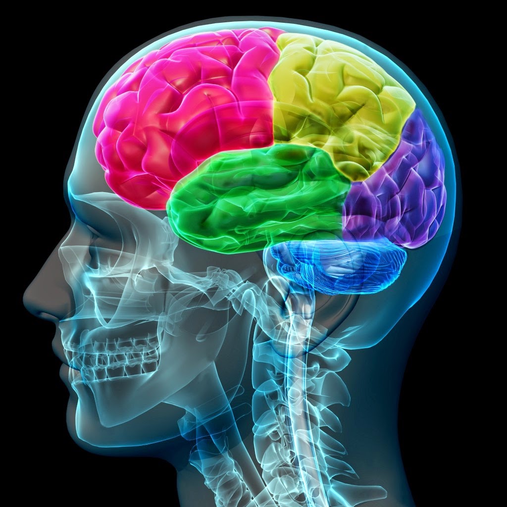
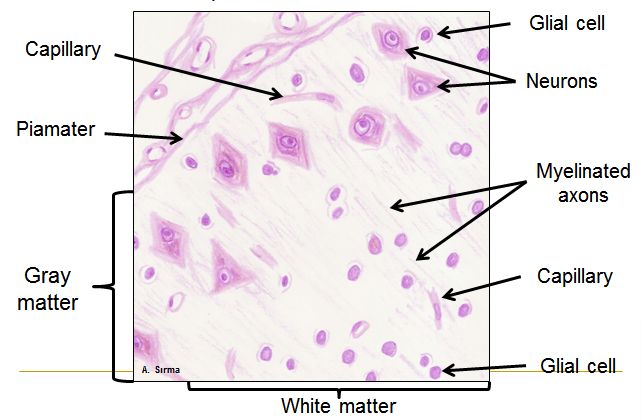
The human brain weighs about 1.4 kilograms and contains around 86 billion neurons. The most noticeable part is the cerebral hemispheres, covered with convoluted cortex. This cortex, or neocortex, is a thin layer of gray matter only 2-4 millimeters thick, but it is where our higher cognitive processes occur.
Neocortex consists of six layers of neurons, each with its own specialization. The first layer contains mainly dendrites and axons running parallel to the cortex surface. The second and third layers contain small pyramidal neurons that form connections within the cortex. The fourth layer is the main “input gate” of the cortex, where axons from the thalamus terminate. The fifth layer contains large pyramidal neurons whose axons go to other cortical areas and subcortical structures. The sixth layer sends signals back to the thalamus.
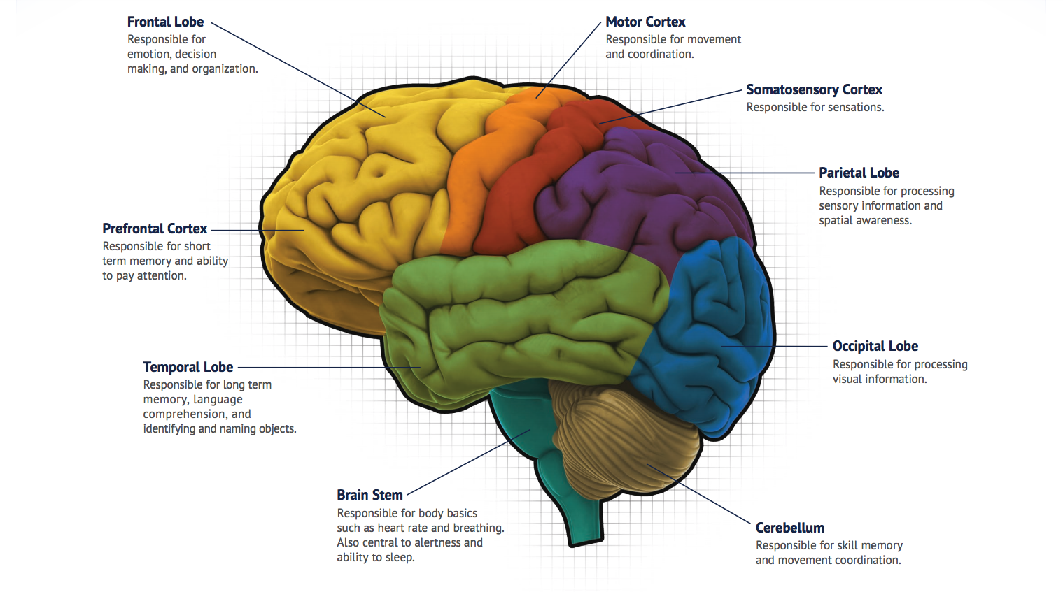
Frontal lobe is our center of executive control. The prefrontal cortex, located in the very front part of the frontal lobe, is responsible for planning, decision-making, working memory, and impulse control. It is this area that makes us human in the full sense — it continues to develop until age 25 and allows us to think about the future, build complex plans, and control our behavior. The premotor cortex plans complex sequences of movements, and the primary motor cortex directly controls muscles through the spinal cord.
Parietal lobe is the center of sensory information integration. The primary somatosensory cortex contains a precise map of the body surface, where lips and hands occupy disproportionately large areas, reflecting their high sensitivity. The posterior parietal cortex integrates information from different senses, creating a coherent representation of the space around us and our body’s position in that space.
Temporal lobes process auditory information and contain structures critical for memory. The primary auditory cortex in the superior temporal gyrus analyzes sounds, and adjacent areas specialize in speech recognition. The inferior temporal cortex contains neurons that selectively respond to faces, allowing us to instantly recognize familiar people in a crowd.
Occipital lobe is almost entirely dedicated to processing visual information. The primary visual cortex receives information from the retina through the lateral geniculate nucleus of the thalamus. Here, primary analysis of visual stimuli occurs: detection of edges, line orientations, motion. Secondary visual areas specialize in more complex tasks: recognition of shapes, colors, moving objects.
Limbic System: The Emotional Brain
Deeper in the brain, beneath the cortex, lies the limbic system — the ancient emotional brain that we share with other mammals. These structures are evolutionarily older than the cortex and control our basic emotions, motivation, and memory.
Hippocampus is our memory center. This curved structure, resembling a seahorse, is critical for forming new memories. Patients with hippocampal damage, like the famous H.M., lose the ability to form new memories.
The hippocampus contains special place cells that activate when an animal is in a specific location in space, creating a cognitive map of the environment.
Amygdala is our center for fear and emotional evaluation. This small structure the size of an almond can instantly assess danger and trigger the “fight or flight” response before the information reaches consciousness. The amygdala also participates in forming emotional memories — that’s why we vividly remember events associated with strong emotions.
Hypothalamus is the link between the nervous and endocrine systems. Despite its tiny size (only 4 grams), it regulates body temperature, hunger, thirst, sexual behavior, circadian rhythms, and hormone production. The suprachiasmatic nucleus of the hypothalamus is our biological clock, synchronizing all processes in the body with the 24-hour cycle.
Basal Ganglia: Center of Movement and Habits
The basal ganglia are a group of interconnected structures deep in the brain that play a key role in movement control and habit formation. The caudate nucleus and putamen together form the striatum — the input gates of the basal ganglia. They receive information from the entire cortex and send it to the globus pallidus, which acts as the main output node of the system.
The substantia nigra contains dopaminergic neurons that modulate striatal activity. Degeneration of these neurons leads to Parkinson’s disease with characteristic motor impairments. The subthalamic nucleus acts as an “emergency brake,” capable of quickly stopping unwanted movements.
The basal ganglia work as a complex system of filters that suppresses unnecessary movements and allows only desired actions to be performed. They are also critically important for learning habits — automatic sequences of actions that do not require conscious control.
Brainstem: Life Support Center
The brainstem is our “reptilian” part that controls vital functions. The medulla oblongata contains centers for respiration and circulation — damage to this area is often fatal. The pons connects different parts of the brain and participates in sleep regulation. The midbrain contains centers controlling eye movements and pupillary reflexes.
The brainstem also houses the reticular formation — a diffuse network of neurons that regulates the level of consciousness and attention. Damage to this system can lead to coma or vegetative state.
Cerebellum: Movement Coordinator
The cerebellum is the “little brain” that contains more neurons than all other parts of the brain combined. It receives copies of all motor commands from the cortex and compares them with sensory feedback, constantly adjusting our movements. The cerebellum also participates in learning motor skills, coordination, and maintaining balance.
Modern research shows that the cerebellum is involved not only in movements but also in cognitive functions — planning, attention, language. There is a close connection between the cerebellum and prefrontal cortex, allowing “tuning” not only movements but also thoughts.
Thalamus: Brain’s Relay Station
The thalamus is an oval structure in the center of the brain that acts as a relay station for most sensory signals going to the cortex. Almost all information from the senses (except olfaction) passes through the thalamus, which filters and processes it before sending to the corresponding cortical areas.
The thalamus consists of many nuclei, each specializing in a certain type of information. The lateral geniculate nucleus processes visual information, the medial geniculate nucleus — auditory, ventroposterior nuclei — somatosensory.
Neurons: Fundamental Units of Information
Neuron Anatomy: Structure for Function
A neuron is a highly specialized cell optimized for receiving, processing, and transmitting information. Each neuron can be divided into four main parts, each performing its unique function.
Soma (cell body) contains the nucleus with DNA and most organelles necessary for cell life. Here, proteins are synthesized, main metabolic processes occur, and “decisions” are made about whether to generate an action potential. The soma size varies from 5 micrometers in small interneurons to 100 micrometers in large motor neurons.
Dendrites are branched processes that receive signals from other neurons. The name comes from the Greek word “tree,” and indeed, dendrites resemble tree branches. In some neurons, the dendritic tree can be very simple — just a few short processes, in others — incredibly complex, with thousands of branches. Pyramidal cortical neurons have two types of dendrites: basal, extending from the base of the soma, and apical, going to the cortex surface.
Dendrites are covered with neurotransmitter receptors and capable of active information processing. They do not just passively receive signals but can amplify or weaken them, integrate information from different sources, and even generate their own local action potentials — dendritic spikes.
Axon is a long process that transmits signals from the soma to other neurons, muscles, or glands. Axons can be incredibly long — from a few micrometers in interneurons to over a meter in motor neurons controlling foot muscles. The axon starts from the axon hillock — a specialized area of the soma with a high concentration of sodium channels, where action potentials are generated.
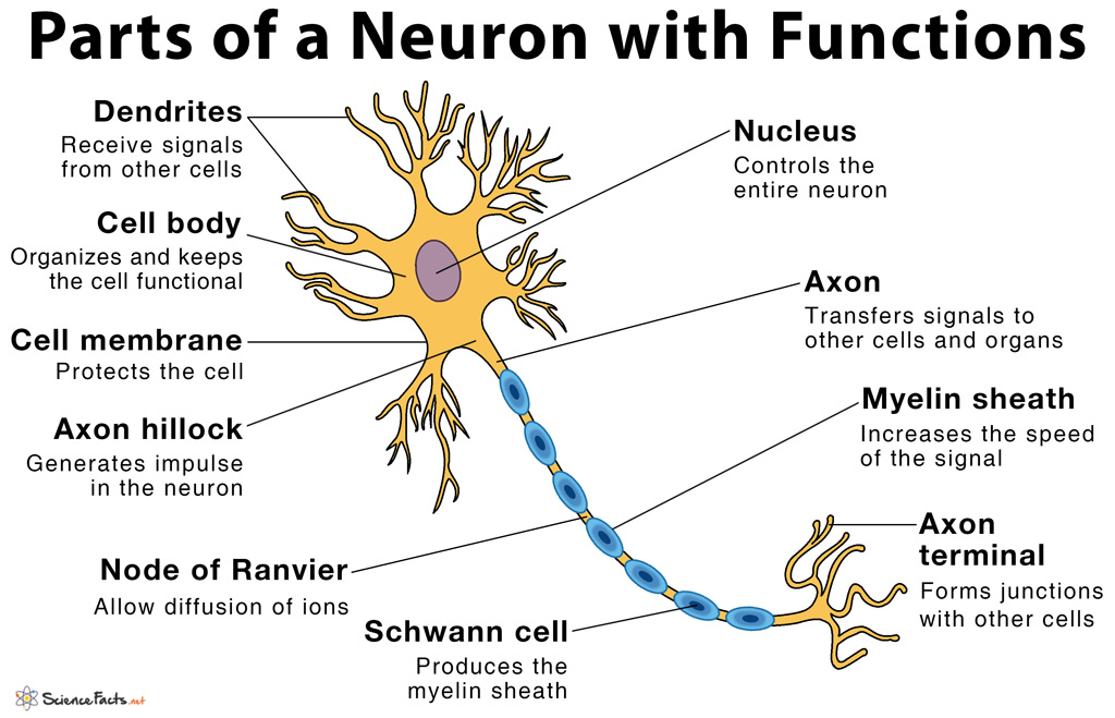
Many axons are surrounded by a myelin sheath — a multilayer membrane formed by glial cells. Myelin acts as insulation, increasing the speed of nerve impulse conduction from 1 m/s to 100 m/s. The myelin sheath is interrupted every 1-2 millimeters at Ranvier nodes, where sodium channels are concentrated. This allows the action potential to “jump” from node to node — saltatory conduction.
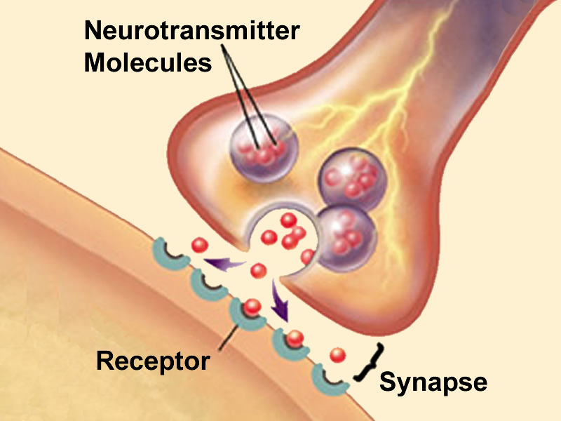
Synapses are specialized contacts between neurons where information is transmitted. The presynaptic terminal contains synaptic vesicles filled with neurotransmitters. When an action potential arrives, the vesicles fuse with the membrane and release their contents into the synaptic cleft, 20-50 nanometers wide. Neurotransmitters bind to receptors on the postsynaptic membrane, causing changes in its electrical properties.
Types of Neurons: Diversity of Forms and Functions
Neurons are incredibly diverse in their morphology and functions. There are several ways to classify them.
By function:
- Sensory neurons receive information from the external environment through specialized receptors. Retinal photoreceptors convert light into electrical signals, skin mechanoreceptors respond to touch and pressure, olfactory and taste chemoreceptors detect molecules.
- Motor neurons transmit commands to muscles and glands. Spinal cord alpha motor neurons directly innervate skeletal muscles, gamma motor neurons control muscle spindles, autonomic neurons regulate internal organs.
- Interneurons make up the vast majority of brain neurons and connect other neurons together. They process information, integrate signals, create complex patterns of activity.
By morphology:
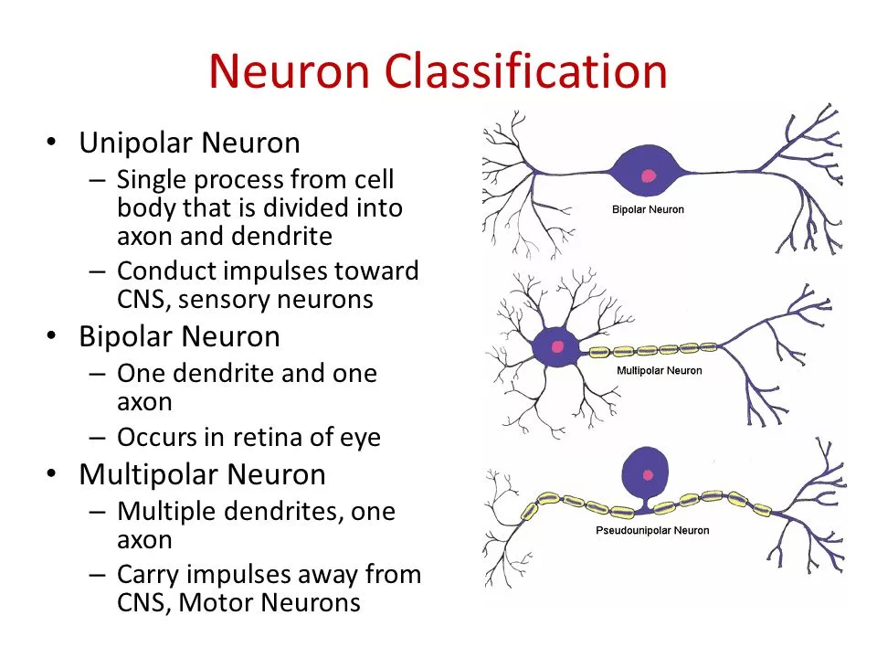
- Unipolar neurons have one process from the soma, which then divides into axon and dendrite. This structure is characteristic of sensory neurons in the peripheral nervous system.
- Bipolar neurons have two processes — one dendrite and one axon, extending from opposite ends of the soma. Found in the retina, olfactory system, vestibular apparatus.
- Multipolar neurons have many dendrites and one axon. This is the most common type in the central nervous system.
By size:
- Granule cells of the cerebellum — the smallest neurons in the brain (5-8 micrometers)
- Pyramidal neurons of the cortex — medium-sized (10-50 micrometers)
- Purkinje cells of the cerebellum and spinal cord motor neurons — the largest neurons (up to 100 micrometers)
By neurotransmitter type:
- Glutamatergic neurons use glutamate and usually have excitatory effects
- GABAergic neurons use GABA and have inhibitory effects
- Dopaminergic neurons modulate the activity of other neurons using dopamine
- Serotonergic, noradrenergic, cholinergic neurons use corresponding neuromodulators
Glial Cells: Unnoticed Helpers
For a long time, glial cells were considered just the “glue” of the brain (from the Greek “glia” — glue), but modern research shows that they play an active role in many brain functions. There are approximately as many glial cells in the brain as neurons, and by volume, they make up about half of the brain matter.
Astrocytes are the most numerous glial cells in the brain. They have a star-shaped form with many processes that contact both neurons and blood vessels. Astrocytes perform many critically important functions:
- Structural support for neurons
- Regulation of ionic composition of the extracellular environment
- Uptake of excess neurotransmitters (especially glutamate)
- Participation in the formation of the blood-brain barrier
- Supplying neurons with nutrients
- Participation in synaptic transmission and plasticity
Oligodendrocytes in the central nervous system and Schwann cells in the peripheral nervous system form myelin sheaths around axons. One oligodendrocyte can myelinate up to 40 axons, while a Schwann cell myelinates only one axon segment.
Microglia are the immune cells of the brain, derived from macrophages. They constantly patrol brain tissue, detecting damage, infections, or dead cells. When activated, microglia can phagocytose pathogens and cellular debris, release anti-inflammatory or pro-inflammatory substances.
Ependymal cells line the brain ventricles and the central canal of the spinal cord. They participate in the production and circulation of cerebrospinal fluid.
Neurotransmitters: The Chemical Language of the Brain
Synaptic Transmission: From Electricity to Chemistry and Back
Imagine two neurons as two people who need to transmit a message across a river where shouting is not possible. The first person (sending neuron) must send a boat (chemical signal) to the second (receiving neuron). This entire process occurs in the synapse.
- The action potential reaches the presynaptic terminal
The action potential is simply a nerve impulse, a short electrical spark that runs along the long process of the neuron (axon). The presynaptic terminal is the very end of this process, a kind of “dock” on the riverbank from which the message will be sent.
Analogy: The first person (neuron) runs to the edge of their bank (terminal), carrying an important assignment (electrical impulse).
- Depolarization opens voltage-gated calcium channels
When the electrical impulse reaches the “dock,” it changes the electrical charge on its surface. This change is called depolarization. In response to this change, special gates on the “dock” open — voltage-gated calcium channels. They are called so because they “depend” on the change in electrical “potential” (voltage) to open.
Analogy: Upon arrival, the person presses a lever (depolarization), which opens the sluice gates in the dam (calcium channels).
- Calcium influx triggers synaptic vesicle fusion with the membrane
There are always many calcium ions outside the neuron. As soon as the gates open, calcium rushes inside the neuron, like water through an open sluice. This flow of calcium is the key signal to action. It causes synaptic vesicles (tiny bubbles filled with chemical “messages”) to move to the very edge of the “dock.”
Analogy: The water gushing from the sluices (calcium) pushes the boats (vesicles) that were ready on the bank toward the edge.
- Exocytosis releases neurotransmitter into the synaptic cleft
Exocytosis is the process when vesicle bubbles fuse with the outer shell of the neuron and “spit out” their contents outside. This content is the neurotransmitter — the chemical mediator, the very “letter” in our analogy. It enters the synaptic cleft — a narrow space between two neurons, our “river.”
Analogy: The boats (vesicles) reach the edge of the bank, fuse with it, and from them, parcels (neurotransmitters) are dumped into the river (synaptic cleft).
- Neurotransmitter diffusion through the synaptic cleft
Diffusion is a natural process in which molecules move from an area where there are many of them to where there are few. The released neurotransmitter molecules simply passively “swim” across the narrow synaptic cleft to the other bank.
Analogy: The parcels dumped into the river are carried by the current to the other bank.
- Binding to postsynaptic membrane receptors
On the surface of the receiving neuron (postsynaptic membrane), there are receptors. These are special protein structures that perfectly fit a specific neurotransmitter in shape, like a lock to a key. When the neurotransmitter “reaches,” it inserts into its receptor.
Analogy: On the other bank, the person (receiving neuron) holds special boxes with locks (receptors), and each parcel (neurotransmitter) perfectly fits its lock.
- Receptor activation changes membrane permeability
As soon as the “key is inserted into the lock,” the receptor activates and changes its shape. This, in turn, opens other channels already on the receiving neuron. As a result, the membrane permeability changes — it begins to allow other ions inside (for example, sodium).
Analogy: As soon as the parcel is inserted into the box, the box lid opens, and through it, something enters the second person’s house (for example, air or light).
- Postsynaptic potential — local change in membrane potential
The flow of ions into the receiving neuron creates a new, its own small electrical shift — the postsynaptic potential. This is the received message. This signal can be either excitatory (command “Act further!”) or inhibitory (command “Stop!”). If enough such excitatory signals accumulate, the receiving neuron will generate its own full action potential and pass the baton on.
Analogy: Having received enough “light” from the open boxes, the second person decides it’s time to act and runs to pass the message to the next.
- Neurotransmitter inactivation
The message is delivered, but to allow the system to receive a new one, the “river” must be cleared of old “parcels.” Otherwise, they will endlessly stimulate the receptors. There are two main mechanisms for this:
Reuptake: The sending neuron, like a vacuum cleaner, sucks back unused neurotransmitters for reuse.
Enzymatic breakdown: Special protein-enzymes float in the synaptic cleft, which, like scissors, cut neurotransmitter molecules, making them inactive.
Analogy: The first person either catches the remaining parcels from the river with a net to use them again, or special cleaning robots in the river simply disassemble them into parts.
Thus, the electrical signal turns into a chemical one to cross the “river” between neurons, and then becomes electrical again, ensuring fast and accurate information transmission in the brain. 🧠
Major Neurotransmitters and Their Functions
Glutamate — the main excitatory neurotransmitter of the central nervous system. Up to 80% of synapses in the cortex use glutamate. It activates several types of receptors:
- AMPA receptors — fast ionotropic receptors providing basic synaptic transmission
- NMDA receptors — slow receptors requiring simultaneous binding of glutamate and membrane depolarization. Critically important for synaptic plasticity and learning
- Kainate receptors — less studied ionotropic receptors
- Metabotropic receptors — G-protein coupled receptors triggering intracellular cascades
Excess glutamate can be toxic to neurons (excitotoxicity), which occurs in stroke, traumatic brain injuries, and neurodegenerative diseases.
GABA (gamma-aminobutyric acid) — the main inhibitory neurotransmitter of the brain. GABA activates two types of receptors:
- GABA-A receptors — fast ionotropic receptors passing chloride ions and causing hyperpolarization
- GABA-B receptors — slow metabotropic receptors
The balance between glutamatergic and GABAergic transmission is critical for normal brain function. Imbalances are associated with epilepsy, anxiety disorders, schizophrenia.
Dopamine — neurotransmitter of the reward and motivation system. There are four main dopaminergic pathways in the brain:
- Mesolimbic pathway (from ventral tegmental area to nucleus accumbens) — reward system, motivation, addiction formation
- Mesocortical pathway (to prefrontal cortex) — executive functions, working memory
- Nigrostriatal pathway (from substantia nigra to striatum) — movement control
- Tuberoinfundibular pathway — regulation of prolactin secretion
Dopamine deficiency in the nigrostriatal pathway causes Parkinson’s disease, excess in the mesolimbic pathway is associated with schizophrenia and psychoses.
Serotonin (5-hydroxytryptamine) is synthesized from the amino acid tryptophan and regulates mood, sleep, appetite, aggression. Most serotonergic neurons are in the raphe nuclei of the brainstem and project throughout the brain. There are over 14 types of serotonin receptors, explaining the diversity of this neurotransmitter’s effects.
Serotonin deficiency is associated with depression, anxiety disorders, obsessive-compulsive disorder. Most antidepressants (SSRIs) block serotonin reuptake, increasing its concentration in synapses.
Norepinephrine (noradrenaline) is synthesized from dopamine and functions as both a neurotransmitter and hormone. In the brain, noradrenergic neurons are concentrated in the locus coeruleus of the brainstem and project to the cortex, hippocampus, cerebellum. Norepinephrine regulates:
- Level of wakefulness and attention
- Stress responses
- Memory consolidation
- Mood
Acetylcholine — the first discovered neurotransmitter. In the central nervous system, cholinergic neurons are concentrated in several nuclei:
- Basal nucleus of Meynert — projections to the cortex, important for attention and learning
- Septal nuclei — projections to the hippocampus, critically important for memory
- Brainstem nuclei — regulation of sleep-wake cycle
Degeneration of cholinergic neurons is characteristic of Alzheimer’s disease, explaining memory problems in this condition.
Neuromodulators: Fine-Tuning the Brain
Unlike classical neurotransmitters, which cause fast and local changes in postsynaptic neurons, neuromodulators act more slowly and on large populations of neurons, changing their sensitivity to other signals.
Endogenous opioids (endorphins, enkephalins, dynorphins) — natural “drugs” of the brain. They bind to opioid receptors and cause analgesia, euphoria, stress reduction. The endogenous opioid system is activated in pain, stress, physical exercise, sex, food intake.
Endocannabinoids (anandamide, 2-AG) act retrogradely — synthesized in the postsynaptic neuron and affect the presynaptic one. They participate in regulation of appetite, pain, mood, memory. They play a protective role, preventing excessive neuron activation.
Adenosine accumulates during wakefulness and causes sleepiness. Caffeine blocks adenosine receptors, preventing the feeling of fatigue and maintaining wakefulness.
Histamine is synthesized in the hypothalamus and participates in regulation of the sleep-wake cycle, attention, and appetite. Antihistamine drugs often cause drowsiness precisely due to blocking histamine receptors in the brain.
Nitric oxide (NO) — a gaseous neurotransmitter that is not stored in vesicles but synthesized as needed. It participates in long-term potentiation, regulation of brain blood flow, and neurotoxic processes.
Electrical Activity of the Brain: Potentials and Rhythms
Membrane Potential: Basis of Neuronal Activity
Each neuron is a miniature biological battery. At rest, the inside of the neuron is negatively charged relative to the external environment — the resting membrane potential is about -70 millivolts. This potential is maintained by the active work of the sodium-potassium pump, which pumps three sodium ions out in exchange for two potassium ions in, creating an electrochemical gradient.
The neuron membrane contains many ion channels — protein pores that selectively pass certain ions. Leak potassium channels are constantly open and allow potassium to exit the cell along the concentration gradient. Sodium channels are closed at rest, but there are many more outside the cell, and they “strive” to enter inside.
Action Potential: Digital Signal of the Brain
When a stimulus is strong enough to depolarize the membrane to a critical level (activation threshold, about -55 mV), a dramatic event occurs — generation of an action potential.
Depolarization phase: Voltage-gated sodium channels open, sodium rushes into the cell in an avalanche, the membrane potential rapidly rises to +40 mV.
Repolarization phase: Sodium channels inactivate, potassium channels open, potassium exits the cell, restoring the negative charge.
Hyperpolarization phase: Potassium channels remain open longer than necessary, the potential becomes more negative than at rest.
Refractory period: For several milliseconds, the neuron cannot generate a new action potential.
The action potential is an “all or nothing” event. It either occurs fully or not at all. Information is encoded not by the amplitude of the action potential (it is always the same), but by frequency — the number of spikes per unit time.
Synaptic Integration: Making Neuronal “Decisions”
Each neuron receives thousands of synaptic inputs from other neurons. Some are excitatory (create EPSPs — excitatory postsynaptic potentials), others inhibitory (create IPSPs — inhibitory postsynaptic potentials). The neuron constantly integrates these signals:
Spatial summation: Simultaneous signals from different synapses add up algebraically.
Temporal summation: Rapidly successive signals from one synapse can accumulate.
If the total depolarization reaches the threshold at the axon hillock, an action potential is generated. Thus, each neuron makes a “decision” based on the balance of all incoming signals.
Brain Rhythms: Symphony of Neuronal Activity
The brain generates rhythmic oscillations of electrical activity that can be recorded using electroencephalography (EEG). These rhythms reflect synchronized activity of large populations of neurons.
Delta rhythm (0.5-4 Hz) — the slowest waves, dominating during deep sleep. They are associated with restorative processes and memory consolidation.
Theta rhythm (4-8 Hz) — characteristic of states of drowsiness, meditation, creative thinking. In the hippocampus, theta rhythm plays an important role in navigation and memory formation.
Alpha rhythm (8-12 Hz) — dominates in a state of calm wakefulness with closed eyes. Reflects the “default mode” of the cortex.
Beta rhythm (12-30 Hz) — associated with active wakefulness, concentration, problem-solving. Excessive beta activity may indicate anxiety or stress.
Gamma rhythm (30-100 Hz) — the fastest oscillations, associated with conscious perception, integration of information from different senses, moments of “insight.”
Neural Networks and Connectome
The brain can be represented as a complex network of 86 billion nodes (neurons) connected by trillions of links (synapses). Connectome is the complete map of all neuronal connections in the brain, analogous to the genome for neural networks.
Local networks: Neurons unite into functional modules — cortical columns, subcortical nuclei, hippocampal layers. Within these modules, connections are particularly dense.
Long-range connections: Long axons connect distant brain areas. The corpus callosum connects the left and right hemispheres, projection fibers connect the cortex with subcortical structures, association fibers connect different cortical areas.
Small-world principle: The brain is organized as a “small-world” network — most neurons are connected to nearest neighbors, but there are also long-range connections that allow information to spread quickly throughout the network.
Hubs: Some brain areas act as nodal stations with particularly many connections. Such hubs include the prefrontal cortex, cingulate cortex, precuneus. Damage to hubs can have catastrophic consequences for brain functions.
Brain Plasticity: Ability to Change
Synaptic Plasticity: Basis of Learning and Memory
One of the most remarkable properties of the brain is its ability to change in response to experience. Synaptic plasticity is the change in the strength of connections between neurons, which underlies learning and memory.
Long-term potentiation (LTP) — persistent strengthening of synaptic transmission that can last hours, days, or even years. LTP was first discovered in the hippocampus and follows Hebb’s rule: “neurons that fire together wire together.” The LTP mechanism includes:
- Simultaneous activation of pre- and postsynaptic neurons
- Activation of NMDA receptors and calcium influx
- Activation of intracellular kinases
- Increase in the number of AMPA receptors in the synapse
- Structural changes in the synapse
Long-term depression (LTD) — the opposite process, weakening of synaptic connections. LTD is important for “forgetting” unnecessary information and preventing synapse saturation.
Homeostatic plasticity maintains the overall level of neuron activity in the optimal range, preventing excessive excitation or suppression.
Structural Plasticity: Physical Remodeling of the Brain
The brain can change not only functionally but also structurally. Structural plasticity includes:
Change in dendritic spines: These tiny protrusions on dendrites contain most excitatory synapses. Spines can appear, disappear, and change shape within minutes or hours. Learning new skills increases spine density in corresponding cortical areas.
Axon and dendrite growth: Neurons can elongate their processes, forming new connections. This process is particularly active in early development but continues in the adult brain.
Neurogenesis: It was long thought that new neurons do not form in the adult brain, but research has shown that neurogenesis occurs in the hippocampus and possibly in some other areas. Physical exercise, learning, and enriched environment stimulate neurogenesis.
Gliogenesis: Formation of new glial cells occurs more actively than neurogenesis. New oligodendrocytes can myelinate previously unmyelinated axons, accelerating signal conduction.
Critical Periods of Development
Brain plasticity is maximal in early development when basic neural networks are formed. Critical periods are temporal windows when the brain is particularly sensitive to certain types of experience.
Visual system: If a kitten does not receive normal visual experience in the first weeks of life, the visual cortex does not develop normally, leading to permanent vision impairments.
Language: There is a critical period for language acquisition that ends in adolescence. Children deprived of language interaction in early age may never fully master language.
Musical ear: Absolute pitch develops only with early musical training, usually before 6-7 years.
Compensatory Plasticity
The brain has an amazing ability to compensate for damage. Cross-modal plasticity allows brain areas deprived of usual inputs to reorganize for processing information from other senses. In the blind, the visual cortex can participate in processing tactile and auditory information, explaining their heightened abilities in these areas.
Vicarious functions — the ability of undamaged brain areas to take over the functions of damaged ones. This is especially pronounced in the child’s brain — children can recover from serious brain damage better than adults.
Memory: Architecture of Human Experience
Multilevel Model of Memory
Memory is not a single process but a complex system of interacting components. Atkinson-Shiffrin model identifies three main levels:
Sensory memory — ultra-short storage of sensory information (0.5-3 seconds). Iconic memory preserves visual images, echoic memory — sounds. Most sensory information is quickly forgotten if it does not attract attention.
Short-term memory stores a limited amount of information (7±2 items) for seconds or minutes. Information is maintained by active rehearsal and can be transferred to long-term memory.
Working memory — an active form of short-term memory that not only stores but also manipulates information. Baddeley’s model includes:
- Phonological loop for verbal information
- Visuospatial sketchpad for visual information
- Central executive for attention control
- Episodic buffer for information integration
Long-term memory has virtually unlimited capacity and can retain information for decades.
Types of Long-Term Memory
Declarative (explicit) memory — conscious recollections:
- Episodic memory — memories of personal events tied to time and place (“what,” “where,” “when”)
- Semantic memory — knowledge about the world, facts, concepts not tied to specific events
Non-declarative (implicit) memory — unconscious forms of memory:
- Procedural memory — skills and habits (riding a bike, playing an instrument)
- Priming — influence of previous experience on current perception
- Classical conditioning — associative links between stimuli
- Non-associative learning — habituation and sensitization
Neurobiology of Memory
Hippocampus — central structure for forming new declarative memories. It does not store memories permanently but is necessary for their consolidation — the process of transferring from short-term to long-term memory. Patient H.M., who had his hippocampus removed for epilepsy treatment, lost the ability to form new memories but retained old ones.
Entorhinal cortex — “gate” to the hippocampus, contains grid cells that create a spatial coordinate system.
Neocortex — site of long-term memory storage. Different types of information are stored in corresponding areas: visual memories in visual cortex, auditory in auditory cortex.
Amygdala enhances consolidation of emotionally significant memories, explaining why we better remember events associated with strong emotions.
Basal ganglia are critically important for procedural memory and habit formation.
Memory Processes
Encoding — the process of translating information into a form suitable for storage. Encoding efficiency depends on:
- Depth of processing (semantic processing is better than superficial)
- Material organization
- Linking with existing knowledge
- Emotional significance
Consolidation occurs in two stages:
- Synaptic consolidation (minutes-hours) — stabilization of synaptic changes
- Systems consolidation (months-years) — gradual transfer of memories from hippocampus to neocortex
Retrieval — the process of reproducing stored information. Each retrieval can change the memory (reconsolidation), explaining why memories are not always accurate.
Forgetting — not just passive loss of information but an active process. Forgetting can be adaptive, allowing the brain to discard unnecessary information and focus on the important.
False Memories and the Reconstructive Nature of Memory
One of the most striking discoveries in neurobiology is that memory does not work like a video recording. Every time we recall something, the brain reconstructs the memory anew, using fragments of information and filling gaps based on our knowledge, beliefs, and current context.
False memories can seem as vivid and convincing as real ones. Elizabeth Loftus’s experiments showed how easily false memories of events that never happened can be implanted. People can “recall” details about getting lost in a shopping mall as a child, even though it never happened.
Source monitoring effect explains why we can remember information but forget where it came from. A familiar phrase may seem like our own thought, a quote from a book — personal experience. This effect underlies many cognitive biases and explains how false beliefs spread.
Cryptomnesia — unintentional plagiarism when others’ ideas are mistakenly perceived as one’s own. This happens because memory for content and memory for source are processed by different neural systems.
Attention: Spotlight of Consciousness
Architecture of Attention
Attention is not a single process but a family of related mechanisms that allow the brain to selectively process information. Michael Posner identified three main attention networks:
Alerting Network maintains readiness to perceive stimuli. It is activated by noradrenergic projections from the locus coeruleus and ensures general wakefulness level. Disruptions in this network are associated with reduced concentration and attention deficit.
Orienting Network directs attention in space. It includes:
- Superior parietal cortex — controls voluntary attention shifts
- Temporoparietal junction — responds to unexpected stimuli
- Frontal eye fields — coordinate attention and eye movements
Executive Network resolves conflicts between competing processes. It is centered in the anterior cingulate cortex and dorsolateral prefrontal cortex. This network is activated when it is necessary to suppress automatic responses or switch between tasks.
Selective Attention: Filter of Reality
The brain is constantly bombarded with a huge amount of sensory information — millions of bits per second. Consciousness can process only a tiny fraction of this stream. Selective attention acts as a filter, determining which information will reach consciousness.
Cocktail party effect demonstrates how we can focus on one conversation in a noisy room, ignoring other sounds. But if someone mentions our name in another conversation, attention instantly switches. This shows that the brain continuously monitors all incoming information, even that which is not conscious.
Change blindness shows the limitations of our attention. People may not notice large changes in a visual scene if their attention is directed elsewhere. In the famous invisible gorilla experiment, half of the subjects did not notice a person in a gorilla suit walking through a basketball court when they were counting ball passes.
Inattentional blindness — a metacognitive illusion in which people overestimate their ability to notice changes. We think our perception is full and detailed, although in reality it is extremely selective.
Types of Attention
Sustained attention — the ability to maintain focus on a task for a long time. It is critically important for learning and productive work but requires constant effort, as the brain naturally tends to wander.
Divided attention — attempting to perform multiple tasks simultaneously. Contrary to the popular myth of multitasking, the brain cannot effectively process multiple streams of complex information at once. What we call multitasking is actually rapid switching of attention between tasks, which reduces performance and increases errors.
Covert attention — directing attention without eye or head movements. We can mentally focus on the periphery of vision while continuing to look straight ahead. This is an evolutionarily important ability — a predator can covertly observe prey without revealing intentions.
Neuronal Mechanisms of Attention
Attention changes neuron activity in several ways:
Signal enhancement: Neurons processing important information increase firing rates. If attention is directed to a certain area of the visual field, visual cortex neurons whose receptive fields are in that area become more active.
Synchronization: Attention increases the synchrony of discharges between neurons, improving information transmission. Gamma rhythm (30-100 Hz) is particularly important for binding information from different brain areas.
Noise suppression: Attention suppresses activity of neurons processing irrelevant information, improving signal-to-noise ratio.
Top-down control: Prefrontal cortex sends control signals to sensory areas, determining which stimuli will be processed with priority.
Emotions: Chemistry of Feelings
Functions of Emotions
Emotions are not just subjective experiences but complex adaptive programs that coordinate physiological, behavioral, and cognitive responses to significant events. They perform several critically important functions:
Motivational function: Emotions direct behavior, making us approach beneficial stimuli (joy, interest) and avoid harmful ones (fear, disgust).
Communicative function: Emotional expressions convey information about our intentions and states to others, facilitating social interaction.
Cognitive function: Emotions influence attention, memory, and decision-making. Positive emotions broaden attention focus and stimulate creative thinking, negative ones narrow focus and activate analytical thinking.
Regulatory function: Emotions help adapt to changing conditions, mobilizing the body’s resources for adequate response.
Theories of Emotions
James-Lange theory asserts that emotions are awareness of bodily changes. We do not run because we are afraid; we are afraid because we run. Modern research partially supports this theory — interoception (perception of internal body signals) does influence emotional experiences.
Cannon-Bard theory suggests that emotional experience and physiological arousal occur simultaneously and independently.
Schachter-Singer two-factor theory states that emotion = physiological arousal + cognitive interpretation. The same arousal can lead to different emotions depending on the context.
Ekman’s basic emotions theory identifies several innate, universal emotions: joy, sadness, anger, fear, surprise, disgust. Each has a characteristic facial expression recognized by representatives of all cultures.
Lisa Feldman Barrett’s constructed emotions theory suggests that emotions are not innate programs but constructed by the brain based on previous experience, cultural knowledge, and current context.
Neurobiology of Emotions
Amygdala — central structure for processing emotions, especially fear. It receives sensory information via two paths:
- Fast path: thalamus → amygdala (12 ms) — rough, fast danger assessment
- Slow path: thalamus → cortex → amygdala (40 ms) — detailed stimulus analysis
The amygdala can trigger a fear response before we become aware of the danger. It also forms emotional memories, especially those related to fear.
Hypothalamus regulates physiological aspects of emotions through the autonomic nervous system and endocrine glands. It controls the release of stress hormones, changes in heart rate, breathing, sweating.
Anterior cingulate cortex participates in emotional regulation and decision-making. It evaluates the emotional significance of events and resolves conflicts between emotional and rational impulses.
Insular cortex integrates interoceptive information (signals from internal organs) with emotional experiences. People with more developed insular cortex better awareness of their emotions.
Orbitofrontal cortex evaluates the emotional valence of stimuli and participates in decision-making. Damage to this area (as in Phineas Gage’s case) leads to impairments in emotional regulation and social behavior.
Emotional Regulation
The ability to regulate emotions is a key skill for mental health and social functioning. There are several strategies for emotional regulation:
Cognitive reappraisal — changing the interpretation of a situation to alter the emotional response. This is the most effective and healthy strategy, which activates the prefrontal cortex and suppresses amygdala activity.
Expression suppression — hiding external manifestations of emotions. This strategy requires many resources and can be harmful with constant use.
Attention distraction — shifting attention focus from the emotion-eliciting stimulus to something neutral or positive.
Acceptance — non-judgmental awareness of emotional experiences without attempting to change them. Paradoxically, acceptance often leads to a natural reduction in emotion intensity.
Sleep and Dreams: Neurobiology of Rest
Sleep Architecture
Sleep is not a passive state of rest but an active process critical for physical and mental health. During the night, the brain goes through several sleep cycles, each lasting about 90 minutes.
Non-REM sleep (NREM) is divided into three stages:
Stage 1 (N1) — transition from wakefulness to sleep. Muscle tone decreases, slow eye movements appear, theta waves (4-8 Hz) dominate. Easy to wake up.
Stage 2 (N2) — light sleep. Sleep spindles (12-14 Hz) and K-complexes appear on EEG — characteristic sleep graphoelements. Makes up 45-55% of total sleep time.
Stage 3 (N3) — deep sleep or slow-wave sleep. Delta waves (0.5-4 Hz) dominate. Hard to wake up, upon awakening the person feels disoriented. Critical for physical recovery and memory consolidation.
REM sleep is characterized by rapid eye movements, muscle atonia (except diaphragm), brain activity similar to wakefulness, and vivid dreams. Makes up 20-25% of sleep in adults.
Neurochemistry of Sleep
Sleep and wakefulness are regulated by complex interactions of neurotransmitters and hormones:
Adenosine — “fatigue molecule.” Accumulates during wakefulness as a byproduct of energy metabolism. Binds to adenosine receptors and causes sleepiness. Caffeine blocks these receptors, preventing sleepiness.
Melatonin is synthesized in the pineal gland in response to darkness. It does not cause sleep directly but signals the body about the onset of night and prepares for sleep.
GABA — main inhibitory neurotransmitter, promoting sleep onset. Many sleeping pills enhance GABAergic transmission.
Acetylcholine is released by brainstem neurons during REM sleep and necessary for generating dreams.
Norepinephrine, serotonin, and histamine maintain wakefulness. Their activity decreases during sleep, especially REM sleep.
Functions of Sleep
Memory consolidation: During sleep, information is transferred from short-term to long-term memory. Slow-wave sleep is especially important for consolidating declarative memories, REM sleep — for procedural skills and emotional memories.
Brain clearance: The glymphatic system — the brain’s lymphatic system — is most active during sleep. It flushes out toxic proteins, including beta-amyloid, whose accumulation is associated with Alzheimer’s disease.
Synaptic homeostasis: The synaptic homeostasis hypothesis suggests that sleep weakens synaptic connections that strengthened during wakefulness, preventing synapse saturation and maintaining learning capacity.
Emotional regulation: REM sleep helps process emotional events of the day, integrate them into the existing knowledge system, and reduce their emotional intensity.
Dreams: Theater of the Sleeping Mind
Dreams are one of the most mysterious phenomena of consciousness. Most vivid dreams occur during REM sleep, but dreams can occur in other stages as well.
Activation-synthesis theory by Hobson and McCarley suggests that dreams are the cortex’s attempt to interpret random signals from the brainstem. However, modern research shows that brain activity during REM sleep is not random.
Threat simulation theory suggests that dreams evolved to practice responses to dangerous situations in a safe environment.
Emotional regulation theory views dreams as a way to process emotional conflicts and traumas.
Domhoff’s neurocognitive theory asserts that dreams reflect the same cognitive processes as waking consciousness but in an altered brain state.
Decision-Making: Computational Machine of Choice
Neuroeconomics of Decisions
Decision-making is the process of choosing between different alternatives based on their expected value. The brain constantly solves computational tasks: which food to choose, where to direct attention, whom to interact with.
Reward system is centered around dopaminergic pathways, especially the mesolimbic pathway from the ventral tegmental area to the nucleus accumbens. Dopamine does not encode pleasure directly but signals reward prediction error — the difference between expected and received outcome.
Orbitofrontal cortex encodes the subjective value of different choice options, integrating information about potential rewards and punishments. Neurons in this area respond to abstract value regardless of the specific stimulus modality.
Anterior cingulate cortex tracks conflicts between different choice options and signals the need for cognitive control.
Heuristics and Cognitive Biases
Human decision-making is often irrational from the perspective of classical economic theory. We use heuristics — mental shortcuts that allow quick decisions but can lead to systematic errors.
Availability heuristic: We assess the probability of an event by how easily we can recall examples. This leads to overestimation of rare but vivid events (plane crashes) and underestimation of frequent but unnoticeable ones (heart disease).
Representativeness heuristic: We judge an object’s membership in a category by its similarity to a typical member of that category, ignoring base rates.
Anchoring effect: The first received information disproportionately influences subsequent judgments, even if irrelevant.
Loss aversion: Losses are experienced as more painful than equivalent gains are pleasurable. This explains many irrational economic decisions.
Endowment effect: We value things more when they belong to us, even if we received them randomly.
Dual Systems of Thinking
Daniel Kahneman described two systems of thinking:
System 1 — fast, automatic, intuitive. It operates without conscious effort and is based on associations, emotions, and heuristics. Evolutionarily ancient, energy-efficient.
System 2 — slow, controlled, analytical. Requires conscious effort and attention. Can override System 1 impulses but is resource-intensive and quickly fatigues.
Most of our decisions are made by System 1, and System 2 engages only when necessary. This explains why we often act irrationally, especially under stress or fatigue.
Emotions in Decision-Making
Contrary to the traditional opposition of reason and emotions, modern research shows that emotions are critically important for making good decisions.
Somatic marker (Damasio’s hypothesis) — emotional reactions to possible decision outcomes that guide choice before conscious analysis. Patients with orbitofrontal cortex damage lose the ability for emotional anticipation and make catastrophically bad decisions despite preserved intelligence.
Affect as information: Current emotional state influences evaluation of future events. In good mood, we overestimate positive outcomes, in bad — negative.
Emotional contagion: Others’ emotions influence our decisions through mimicry and empathy. This is especially important in social and economic contexts.
Consciousness: The Last Mystery of the Brain
Definition of Consciousness
Consciousness is perhaps the biggest mystery of science. Why do we have subjective experiences? Why does something feel like something? This is called the “hard problem of consciousness” (David Chalmers).
Phenomenal consciousness — subjective qualities of experience (qualia). Why does red look exactly that way and not another? Why does pain feel exactly that way?
Access consciousness — information available for report, reasoning, behavior control. This is what is usually measured in experiments.
Self-consciousness — awareness of oneself as a separate entity, ability for reflection and metacognition.
Theories of Consciousness
Global workspace theory (Bernard Baars, Stanislas Dehaene) suggests that consciousness arises when information becomes globally available to various brain systems through a network of long cortical connections.
Integrated information theory (Giulio Tononi) asserts that consciousness corresponds to integrated information (Φ) in a system. A system is conscious to the extent that it integrates information in a way inaccessible to its parts.
Predictive brain (Anil Seth, Jakob Hohwy) views consciousness as a controlled hallucination — a model of the world constantly updated based on predictions and errors.
Attention schema theory (Michael Graziano) suggests that consciousness is a simplified model of attention processes that the brain creates for better attention control.
Neural Correlates of Consciousness
Binocular rivalry: When different images are shown to each eye, consciousness alternates between them. This allows studying which brain areas correlate with conscious perception.
Masking: A briefly presented stimulus followed by a mask may not be conscious, although it causes neural activity. Comparing conscious and unconscious stimuli reveals neural correlates of consciousness.
Attentional blink: In rapid presentation of stimulus sequences, the second target stimulus may not be conscious if it follows shortly after the first. This shows temporal limitations of consciousness.
P300 — an evoked potential component that correlates with conscious perception of stimuli. It reflects global spread of information across the cortex.
Gamma synchronization (30-100 Hz) is associated with binding information from different brain areas into a single conscious experience.
Altered States of Consciousness
Anesthesia: General anesthetics suppress consciousness by disrupting connections between different brain areas. Studying the transition from consciousness to unconscious state and back provides important clues about consciousness mechanisms.
Sleep: Transition from wakefulness to sleep is accompanied by changes in brain network connectivity. REM sleep is particularly interesting as it retains many features of conscious experience (dreams) with altered neurochemistry.
Psychedelics: Drugs like LSD, psilocybin alter consciousness by disrupting normal hierarchical brain organization and increasing connectivity between usually separated areas.
Meditation: Long-term meditative practice changes brain structure and function, especially the default mode network associated with self-referential thinking.
Practical Conclusions: How to Use the Brain
Understanding how the brain works gives us several important practical conclusions:
-
Remember your cognitive limitations. Your brain is a device, and it can make mistakes. What seems obvious may be just a feature of how your brain builds a model of the world.
-
Develop rational thinking skills. Since we can sometimes consciously influence our model of the world, it is important to do so carefully and reasonably.
-
Learn constantly. The brain is plastic throughout life. Regular learning and solving new tasks keeps it in good condition.
-
Use proper memorization strategies. Repetition, positive reinforcement, and good functional state are keys to effective learning.
-
Don’t forget about rest. Adequate sleep is important not only for recovery but also for creative processes and memory consolidation.
The brain is not just an organ of thinking; it is a complex machine for creating reality. Understanding its work helps us better know ourselves and more effectively use our cognitive abilities. We are blind informational machines, but awareness of this fact makes us more sighted in navigating the complex world of human cognition.
Related Topics
How to take care of your brain health?
A comprehensive guide to maintaining healthy thinking and mental well-being.

Rationality
About stereotypes regarding rational individuals and what rationality truly entails.